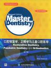
口腔修复学、正畸学与儿童口腔医学
出版时间:2009-1 出版社:北京大学医学出版社 作者:Peter Heasman 页数:359
前言
The philosophy of this textbook remains unchanged from that of the First Edition, where the emphasis was placed on understanding, learning and self-assessment so that the reader is able to explore their own level of knowledge, identify their strengths and, perhaps more importantly, weaknesses or gaps in their knowledge base, which can then be addressed. Basically, the book comprises chapters on aspects of restorative dentistry, paediatric dentistry and orthodontics. There is also a chapter on law and ethics that has been updated considerably since the First Edition as a consequence of the considerable developments and restructuring that have occurred within the General Dental Council of the United Kingdom. Changes with respect to registerable qualifications, development of specialist lists and the International Qualifying Examination have also underpinned significant rewriting of this chapter. There is also a new chapter that addresses the restorative manage- ment of dental implants in which the objective is to pres- ent the reader with the basic restorative concepts of dental implantology whilst recognising that those postgraduates who may have a broadening interest in this subject will need to refer to a more specialist text. Another innovation has been the inclusion of extended matching item (EMI) questions in all of the self-assessment sections. This type of question is becoming more popu- lar as a form of assessment in UK dental schools and it is widely recognised that they are an effective and valid assessment tool as well as being a significant challenge for examiners to prepare! The popularity of various assess- ment tools, however, tends to change on a regular basis and every effort will be made to ensure that the assessment methods presented in this textbook will remain in touch with contemporary education philosophy.Finally, I should like to record my sincerest thanks to the contributing authors to this book, all of whom are recognised experts in their respective specialties and who have worked dilieentlv to update their chapters for this Second Edition.
内容概要
本书是口腔医学专业双语教学的核心教材,以简明易读的形式讲述口腔修复学正畸学与儿童口腔医学基本要点。编写过程中使用了大量示意图与照片,方便学生学习与掌握学科核心内容。每一部分皆附有多种形式的自我测试题和解析,有益于学生复习备考和提高应试能力。本套丛书是口腔医学专业本科生及研究生的实用教材和理想的复习备考用书。
作者简介
作者:(英国)Peter Heasman
书籍目录
ContributorsAcknowledgmentPrefaceUsing this book1.Periodontology Philip Preshaw and Peter Heasman2.Endodontics Philip Lumley3.Conservative dentistry Stewart Barclay4.Prosthodontics Craig Barclay5 Restorative management of dental implants Glies McCracken 6.Conscious sedation in dentistry Nigel Robb7.Paediatric dentistry I Richard Welbury and Alison Cairns8.Paediatric dentistry II Richard Welbury and Alison Caims9.0rthodontics I:development,assessment and treatment planning Declan Millett10.Orthodontics II:management of occlusal problems Declan Millett11.Orthodontics III:appliances and tooth movement Declan Millett12.Law and ethics Douglas LovelockIndex
章节摘录
TechniquesFor effective instrumentation, the operator should be com- fortably seated, the patient should be supine in the dental chair and there should be good illumination. An assis- tant should retract the oral soft tissues, where necessary, and maintain a clean operating field through the use of an aspirator. The operator should have a good knowledge of dental anatomy and root morphology, and appropriately selected, sharp instruments should be used. The modified pen grip is preferred, in which the instrument is held by the thumb, index finger and middle finger in the same way as a pen is held. However, the index finger is bent so it can be positioned well above the middle finger on the instru- ment handle. In this way, a triangle of force is applied to the instrument, and this tripod effect allows for stability and tactility during use. The fourth finger (ring finger) is used as a finger rest to stabilise the hand and reduce the likelihood of uncontrolled movements and injury. The fin- ger rest also acts as a fulcrum for working movements of the instrument. Finger rests may be on tooth surfaces in the immediate vicinity of the working area, they may be cross arch (on tooth surfaces in the other side of the same arch), opposite arch or extraoral.Calculus should be identified prior to RSI. Supragingival calculus can be seen with good lighting and a dry field. The dark colour of subgingival calculus may be visible through thin overlying gingival tissues. An air syringe to retract the gingiva may also reveal subgingival calculus. Interproximal calculus may be visible radiographically. An explorer should be used subgingivally to check for calcu- lus, grooves, furcations and other anatomical structures.Scaling instruments should be adapted to the tooth sur- face, which means that the cutting edge of the instrument conforms to the anatomy of the root surface. This results in maximal efficiency during scaling and minimal damage to the adjacent tissues. The cutting edge should be angled at between 45 and 90 ~~ to the root surface: less than 45° and the instrument will not engage the calculus, more than 90° and tissue trauma will be achieved instead. Instruments should be placed apical to the calculus and pulled coro- nally with firm, controlled strokes to remove the calcu-lus. Increasing lateral pressure may need to be applied to remove particularly tenacious deposits. Following RSI, debris should be flushed from the pocket with an irrigat- ing solution (e.g. chlorhexidine) in a syringe with a blunt needle. Scaling is indicated for removal of plaque and calculus from enamel surfaces. On root surfaces, however, calcu- lus and plaque grow in surface irregularities of cementum, and this thin layer of cementum may need to be removed during RSI. Furthermore, bacterial products, sucl~~ as endo- toxin, penetrate into the cementum surface, and removal of a superficial layer of cementum promotes healing. It is not necessary, however, to remove extensive amounts of cementum and/or dentine during RSI, and to do so can cause dentine hypersensitivity, pulpitis and, at extremes, render the tooth susceptible to fracture.Historically, dentists aimed to leave root surfaces 'glassy smooth and hard' following RSI (referred to then as root planing). However, research has shown that this is not nec- essary, and indeed, may result in excessive damage to the tooth by removal of excess cementum. Instead, we now consider that RSI aims to remove plaque and calculus and disrupt the subgingival biofilm so that the host-parasite balance is tipped in favour of the host to promote healing. The pockets should not be probed sooner than 6-8 weeks after RSI as this may interfere with the healing process. Post-treatment evaluation of the healing response to RSI should be considered together with patient plaque-control capabilities and motivation prior to embarking on further treatments, such as additional RSI, adjunctive antimicro- bial usage or periodontal surgery.
编辑推荐
《口腔修复学、正畸学与儿童口腔医学(第2版)》是口腔医学基本要点丛书之一。
图书封面
评论、评分、阅读与下载
用户评论 (总计13条)
- 我很想通过学习国外的原版教材来看看国外在专业领域的观点,国外的原版书在国内的售价太高了,像我们这样的学生一般都买不起。我觉得这本影印教材非常的好,价格便宜,书中涵盖了基本要点,与临床结合紧密,有大量的图片,是专业学习和专业外语学习的好材料
- 书很不错,很专业!宣称是双语,但是只有英文,何来的双语呢?
- 第一次买书,很方便很满意
- 别人推荐的,看了之后觉得内容很好。
- 当时没认真看,以为是中文的,结果是全英文的,英文水平不是太好,退了。
- 英语不好,慢慢看还是能看懂的,好书
- 慢慢翻译吧。。。
- 不错哦,值得拥有,没有彩页
- 给同学买的,她说挺好的,对专业英语帮助很大,呵呵
- 老师要求买的。是正品吧。
- 太慢了,将近快一个月了。
- 口腔修复学、正畸学与儿童口腔医学(第2版)Peter Heasman到我手里的书很满意
- 特别好,内容好,质量好,都好
相关图书
- 医学英语会话
- 厨房窍门1888例
- 大众菜精选1888例
- H幼儿园活动整合课程托班下故事大书(4)
- 家常主食面点
- 家常凉拌菜
- 精选家常菜
- 速查家常菜
- 常见食材家常菜
- H故事大书大班下(我的幼儿园)5本/套
- H故事大书中班下(同行)5本/套
- I故事大书小班上
- 变色鸟
- D幼儿园早期阅读大书小班上-多多什么都爱吃
- 为什么我不能
- D幼儿园早期阅读大书小班下-阿比比一比
- 收集东 收集西
- D幼儿园早期阅读大书中班上-花园里有什么
- D幼儿园早期阅读大书中班下-彩色的鸭子
- D幼儿园早期阅读大书中班下-白羊村的美容院
- D幼儿园早期阅读大书大班上-没有不方便
- D幼儿园早期阅读大书大班上-国王生病了
- D幼儿园早期阅读大书大班下-棒棒天使
- D幼儿园早期阅读大书大班下-啊哈,幼儿园
- 医药国际贸易
